Before birth
VUR may cause antenatal hydronephrosis which is when one or both of a baby’s kidneys hold on to urine, and become stretched and swollen. This can be seen during pregnancy on an ultrasound scan.
After birth
Some babies with antenatal hydronephrosis or other problems in pregnancy are tested for VUR after birth. Older children may be tested for VUR if they have symptoms of a UTI.
Your child may have a urine test and blood test to check how well their kidneys are working.
They may also have one or more scans. These might include:
Ultrasound scan
A small handheld device is moved around your child’s skin and uses sound waves to create an image on a screen to look at the kidneys and urinary system.
MAG3 scan
A chemical called MAG3 that gives out a small amount of radiation (energy) is injected into one of your child’s blood vessels using a needle or a small plastic tube called a cannula. A local anaesthetic gel or cream can be used to stop your child feeling any pain from the injection. A special camera takes images of your child’s urinary system as the chemical passes through it.
MCUG (micturating cysourethrogram)
A long thin tube called a catheter is inserted into the bladder. A small amount of contrast dye is put through the catheter. A special X-ray machine takes a series of images of the bladder while your child is passing urine. The flow of urine can be seen because of the contrast dye. This test can be used to diagnose VUR.
DMSA scan (Dimercaptosuccinic Acid)
This is a type of radionucleotide scan. This means that a substance that gives out a type of radiation called gamma rays is injected into the blood stream. This substance is taken up by the kidneys and a special camera takes some pictures to show how well the kidneys are functioning and whether there is any damage.
Grading
VUR is categorised into five grades depending on where the reflux happens.
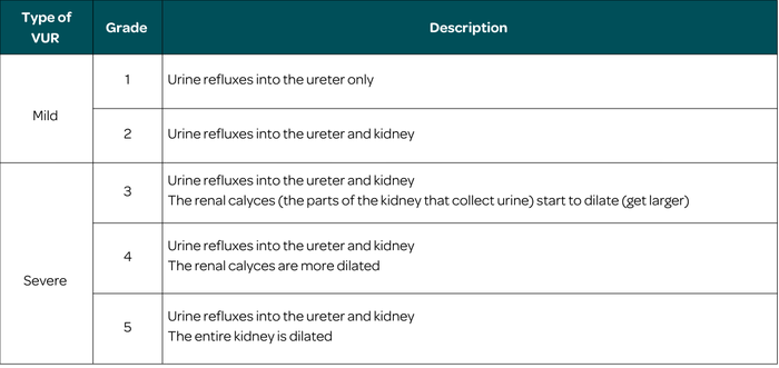
VUR gets better in more than half of children in the first few years of life. Some children stay at the same grade for a long time. A few get worse (go to a higher grade).
This web page is printer-friendly.
To save the page as a PDF, print as normal and select "Save as PDF" as your printer destination.