The urinary system is made up of two kidneys, two ureters, a bladder and a urethra.
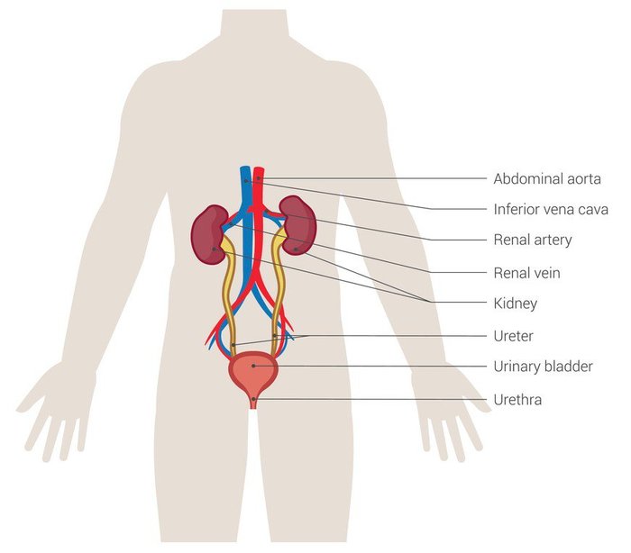
The kidneys are bean-shaped organs. Most people have two kidneys. They sit in your lower back under the bottom ribs.
The two ureters are long tubes that carry urine from the kidneys to the bladder.
The bladder is a round, muscular bag that sits low down in the pelvis and stores urine until it leaves the body when we urinate (wee). The bladder fills up with urine like a balloon. When it is full, it sends signals to the brain to ‘tell’ the bladder muscles to push urine out through the urethra. This is how we pass urine.
The urethra is a tube that carries urine from the bladder to the outside of the body. We urinate through the opening. In boys, the urethra passes through the penis and is much longer than in girls.
What do your child’s kidneys do?
Your child’s kidneys do lots of important jobs to keep them healthy, including:
- controlling your child’s blood pressure
- cleaning their blood.
- helping to keep their bones and teeth strong and healthy by balancing the levels of important minerals in their body.
- removing waste products from their blood by passing them out in their urine and control the amount of salt and water in the body. Your child’s kidneys make up to two litres of urine each day!
- helping to produce red blood cells which carry oxygen around the body.
About the kidneys
A kidney is reddish and shaped like a kidney bean.
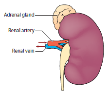
Our kidneys grow as we get older. Each kidney is about the size of our fist.
Most newborn babies have kidneys that are about 4 centimetres long. When they reach eight years old, their kidney will have grown to about 8 centimetres. Adult kidneys are about 13 centimetres long.
We have two adrenal glands, one on top of each kidney. (“Ad” means on top of” and “renal” means to do with the kidney.) They are shaped like pyramids.
The adrenal glands help to control how the kidneys work. They make and release chemicals called hormones, which are carried in the blood to send messages to other parts of the body. These hormones are very important to many of the body’s functions, especially at times of stress such as when we are ill or have an operation.
Like other parts of the body, the kidney needs blood for everything it does. Blood is pumped around the body by the heart, through tubes called blood vessels.
- The renal artery carries blood into the kidney.
- The renal vein carries blood out of the kidney. These large blood vessels go into the kidney at a dip on its side.
Other smaller blood vessels carry blood around the kidney. These have different names, depending on their size. The smallest of these blood vessels are capillaries.
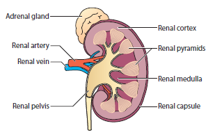
The kidney has a few different areas. Starting from the outside of the kidney, these are:
- renal capsule – a membrane (like a skin) that covers and protects the kidney
- renal cortex – the outer area
- renal medulla – the inner area, which has about eight sections shaped like triangles called renal pyramids
- renal calyces – these are small areas that collect urine made by the kidney
- renal pelvis – the urine flows from the calyces to this larger area before moving down into the ureter, the tube that leads to the bladder.
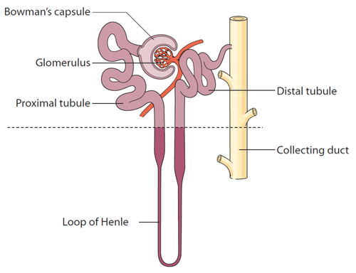
Inside each kidney there are about one million nephrons. Nephrons are tiny but very important units that filter blood and make urine. Each nephron has two main parts:
- glomerulus
- renal tubule
The glomerulus
Blood that comes into the kidney flows into each glomerulus, a tight bunch of capillaries (tiny blood vessels). The glomerulus acts like a sieve – waste and water from the blood pass through it. When we talk about more than one glomerulus, we say glomeruli.
The space that surrounds the blood vessels of the glomerulus is called the Bowman's capsule, or glomerular capsule.
This part of the nephron is in the renal cortex, the outer part of the kidney (above the dotted line in the image).
The renal tubule
The renal tubule is a long, narrow tube, starting from the Bowman’s capsule. Everything that is filtered through the glomerulus goes into the renal tubule. This liquid eventually becomes urine.
Part of the renal tubule is in the renal cortex, and part of it is in the renal medulla.
The tubule has four sections: proximal tubule, loop of Henle, distal tubule and collecting duct.
The wall of the glomerulus works as a filter, letting some substances out and keeping some in. This is called the glomerular filtration membrane. It is quite leaky, and so water and other substances in the blood filter out of the glomerulus. However, blood cells and some proteins are too large to pass through the wall and so stay in the blood vessels.
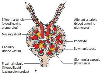
The glomerular filtration membrane is like a sandwich with a few layers.
- Endothelial cells are cells are on the inside surface. They have tiny openings called pores that let small substances through.
- The glomerular basement membrane (GBM) is a layer of material around the endothelial cells.
- Podocytes are cells that wrap around the glomerulus. They have long projections that look like feet – sometimes called foot processes. Liquid can filter through the slits between these “feet”.
- Mesangial cells are cells that sit around the glomerulus and support it. They can contract (tighten) to control how much is filtered through the glomerulus
The liquid that is filtered through the glomerulus flows along the renal tubule in the order shown below.
- Proximal tubule – this is a coiled-up section next to the glomerulus and Bowman’s capsule (proximal means next to).
- Loop of Henle – this is the next part of the renal tubule. It is a U-shaped section that dips down into the renal medulla. Some nephrons have longer loops of Henle.
- Distal tubule – this is a coiled-up section that connects the loop of Henle to the collecting duct. Because it is further away from the glomerulus and Bowman’s capsule, it is called distal (distant or further away).
- Collecting duct – this is the final section of the renal tubule. It is where urine collects before it flows to the renal pelvis before leaving the kidney.
The renal tubule is lined with tubule cells. These cells help carry substances between the renal tubule and the bloodstream.
There are tiny blood vessels around the nephron.
- Blood goes into the glomerulus through the afferent arteriole (afferent means carried in).
- Blood that is not filtered through the glomerulus leaves through the efferent arteriole (efferent means carried out).
- This blood is then passed into the peritubular capillaries, which surround the nephron.
Share this page with a QR code, print or save as PDF
Click on the blue box (on the right or below) and scan the QR code with your phone or tablet.

This web page is printer-friendly.
To save the page as a PDF, print as normal and select "Save as PDF" as your printer destination.